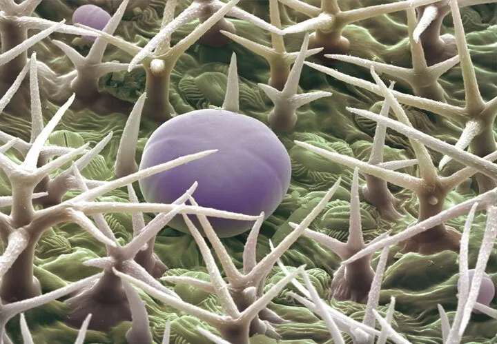
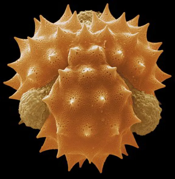
Microscopic Look at Essential Oil

The microscopic images of the different structures in the plant that is responsible for producing the essential oil.

STRUCTURE

A look at the different structures responsible for creating essential oil.

PLANT CHEMISTRY

Plants chemical makeup is important to man's survival. 

INVESTIGATION

Explore how essential oils are stored in a plant. 

Module 2 | Lesson 6

Microscopic Look at Essential Oils

In this lesson, we will look at the plant's different structures responsible for producing the essential oil.


These images carry classical pharmacognosy to a deeper level of detail through the modern technology of scanning electron microscopy. Examining plant structures helps to differentiate between species to assure proper identification and brings a clearer understanding of these benefits and beautiful life forms.


Single pollen grain of chamomile( Matricaria recutita L., Asteraceae) (SEM, critical point dried, magnified 9,313 times actual size).  Photo © Microscopix

Chamomile

Single pollen grain of chamomile(Matricaria recutita L., Asteraceae) (SEM, critical point dried, magnified 9,313 times actual size). Photo © Microscopix photolibrary
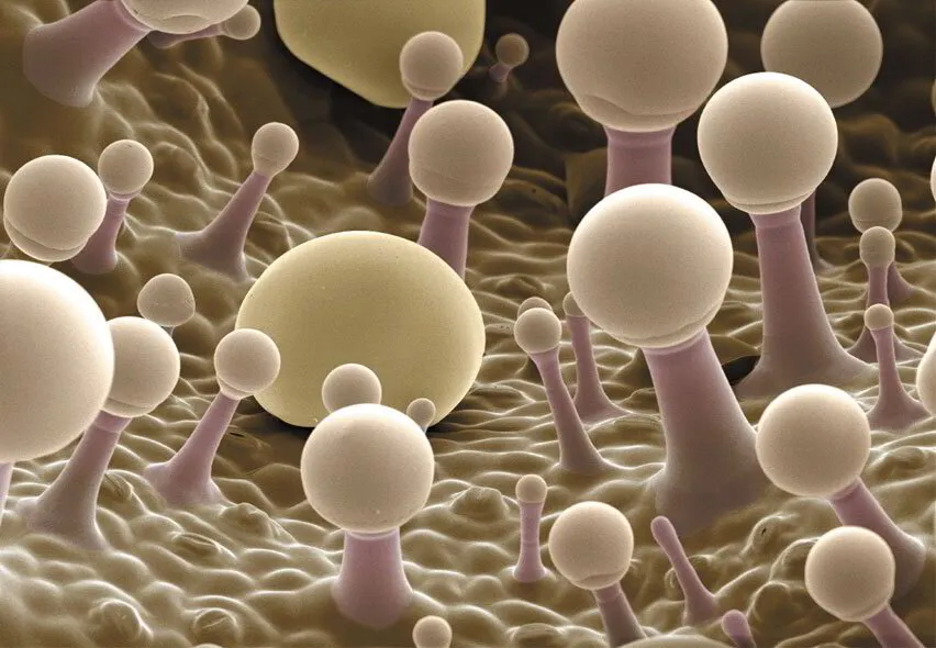
Plant Chemistry

For thousands of years, plants have provided humanity with many of the primary and important materials required for day-to-day living, including oxygen, food, clothing, and timber, as well as being a source of compounds such as oils, resins, rubbers, gums, dyes, pesticides, and drugs.


Plants might be considered biochemical factories that have evolved over the last 440 million years. Researchers elucidate chemical pathways and various industrial applications of plant products, and anatomical description is one of the most important aspects of the economic uses of these plants. Clearly, we should also safeguard and conserve the diversity of our planet’s flora and carefully consider our plant resources’ exploitation.


Plant chemistry became an established university discipline at the end of the 19th century; since then, many new structures have been discovered. The number of natural products obtained from plants exceeds 100,000, and new chemical compounds are discovered each year. Although the functions of some plant chemicals have not been fully established, it is known that some regulate growth. In contrast, others are involved in pollination and seed dispersal or serve as antifungal or defense agents. Still, others are engaged in mutually beneficial (symbiotic) associations with other organisms, such as the partnership between plants that provide food and shelter to bacteria in their roots that convert nitrogen from the air into ammonia fertilizer. These fungi help the plant’s roots to absorb more water and minerals, or ants living in the branches that protect and help nourish the plant.


The complex nature of these chemicals, which are usually produced in various types of secretory structures, is also influenced and controlled by genetic and ecological factors and, significantly, by the mode of extraction from the plants.


The type of secretory structure is an important and differentiating characteristic of many plant families. Detailed anatomical description of these structures is relevant to the market value of the plants, the verification of the authenticity of a given species, and the detection of substitution or adulteration. It also provides a guide to the method of processing.


The microscopic investigation of plant structures is integral to the complex biological research process, including plant growth and development, genetics, and breeding. Observing the endless variability in form and structure with the microscope allows us to discover nature in one of its most powerful forms.

Clary Sage – Salvia sclarea Under Microscope | Aroma Hut Institute

Clary Sage

Clary sage (Salvia sclarea L., Lamiaceae) showing stalked and sessile secretory glands on the calyx trichomes (Cryo-SEM, magnified 752 times actual size). Photo © Microscopix photolibrary
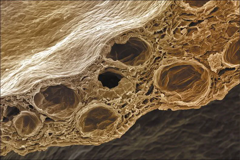
Secretory Structures in Plants

Plant chemicals are classed as primary or secondary metabolites. Primary metabolites are biochemicals that are essential for plant growth and survival. Universally present in all plants, they include sugars, amino acids, nucleic acids, and chlorophylls. Secondary metabolites, from alkaloids to phenols, comprise the remaining plant chemicals and are probably found in all plant species.


Essential oils are complex, volatile mixtures of specific secondary plant metabolites. Many plant species produce essential oils, including annual, biennial, or perennial herbaceous plants, evergreen or deciduous shrubs, and trees.


The ecological and evolutionary role of secondary metabolites has been associated with:


  • Defense against animals
  • Healing of plant organ wounds
  • Protection from harmful insects
  • Resistance to microbial attacks
  • The attraction of insects and animals for pollination


Several species and varieties of plants, primarily those of commercial interest, have been investigated systematically and in-depth. Research groups in university biological and pharmacological departments have recently undertaken various studies concerning secretory structures and factors influencing their development.


A common feature of living cells, secretion involves the discharge of substances to the exterior (exotropia secretion) or into special intercellular cavities (endotrophic secretion). The secreted material may contain various salts, latex, waxes, fats, flavonoids, sugars, gums, mucilages, essential oils, and resins. It has been assumed that these products are biosynthesized in situ, and direct evidence for the biosynthetic capacity of gland cells has become available recently with the development of procedures for gland isolation. These methods have yielded definitive proof of the presence of enzymes within gland cells.


Trichomes (described below) present on the leaf surface and other secretory tissues can be examined using light, scanning, and transmission electron microscopy, which enables detailed observation of major stages in the development of secretory cells, including their membrane system and nuclei, the overall size of the gland, and the amount of material released into the subcuticular cavity.


Essential oils, with or without accompanying resins and gums, are most commonly found in unique secretory structures on the plant's surface or within the plant tissues. The type of structure is specific to the family or species. This can be useful to identify plant material and verify the authenticity of the plant source in the case of suspected adulteration.

Clove Bud - Syzygium aromaticum Under Microscope | Aroma Hut Institute

Clove Bud

The dried petal of clove (Syzygium aromaticum, (L.) Merr. & L.M. Perry, Myrtaceae), showing its many endogenous oil glands (SEM, CPD, magnified 607 times actual size). Photo © Microscopix photolibrary
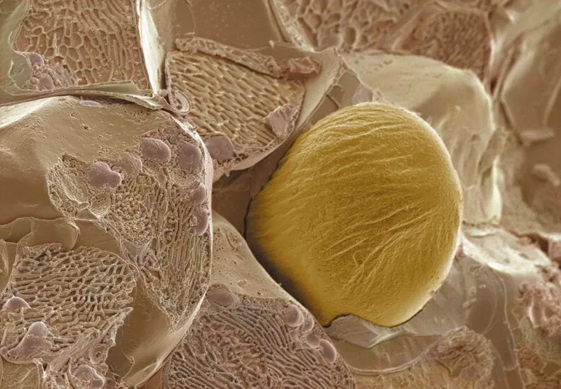
Secretory cells

The simplest secretory structure is a single secretion-containing cell where only the actual content distinguishes it from adjacent non-secretory cells. However, it may also be larger than the other cells or have a thick cuticularized lining. This cell type is found in many different plant tissues, such as in the rhizome pith and cortex of ginger (Zingiber officinale Roscoe, Zingiberaceae) and in the perisperm and embryo of nutmeg (Myristica fragrans Houtt., Myristicaceae).

Ginger Zingiber officinale Under Microscope | Aroma Hut Institute

Ginger

Rhizome of ginger (Zingiber officinaleRoscoe, Zingiberaceae), showing oil globules within the membrane of its secretory cell (Cryo-SEM and etched, magnified 2,149 times actual size). Photo © Microscopix photolibrary
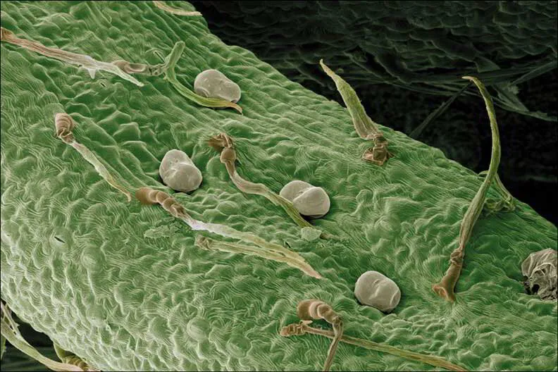
Secretory Cavities

These cavities are more or less spherical structures that can be formed in two ways: the parenchyma cells (i.e., the soft plant tissue composed of thin-walled, relatively undifferentiated cells that may vary in structure and function) can separate one from another, leaving intercellular spaces called lumina or lacuna, or an actual cell can disintegrate and leave a cavity within the tissue.


These spaces are lined with secretory cells, an epithelium, that produces the essential oils. In high oil-yielding plants, several layers of these secretory cells are formed. The cavities continually enlarge, and some become filled with cells with thin, convoluted walls that store the oil produced from within their plastids (a class of cytoplasmic organelles).


This group includes fruits and leaves of plants in the Citrus family (Rutaceae) and Eucalyptus spp. (Myrtaceae). Secretory cavities are also present in the flower buds of cloves (Syzygium aromaticum (L.) Merr. & L.M. Perry, Myrtaceae) and the elongated cavities in the bark of frankincense (Boswellia spp., Burseraceae).

Roman Chamomile - Chamaemelum nobile Under Microscope | Aroma Hut Institute

Roman Chamomile

Upper leaf surface of Roman chamomile(Chamaemelum nobile (L.) All., Asteraceae) with sessile secretory glands and non-secretory trichomes (SEM, CPD, magnified 417 times actual size). Photo © Microscopix photolibrary
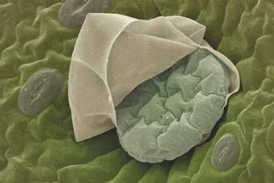
Secretory ducts

Ducts are elongated cavities. They can often branch to create a network extending from the roots through the stem to the leaves, flowers, and fruits. They are composed of an epithelium that surrounds a central cavity. Several predisposed cells within the parenchyma undergo asynchronous division, and in doing so, they expand the initial space in the middle, where the cells are adjacent, to form a cavity. Some of these cells forming the cavity wall will change into secretory epithelial cells.


The oils are biosynthesized within their leucoplasts and move via the endoplasmic reticulum into the cavity. These cavities then become joined to form ducts. They can be found in all of the family Apiaceae (Umbelliferae) as well as the families Asteraceae (Compositae), Clusiaceae (Hypericaceae), and Pinaceae.


Oregano Origanum vulgare Under Microscope | Aroma Hut Institute

Oregano

Sessile secretory gland on lower leaf surface of oregano (Origanum vulgare L., Lamiaceae) with ruptured cuticle revealing individual secretory cells. Nearby stomata are also clearly visible (SEM, CPD magnified 1,979 times actual size). Photo © Microscopix photolibrary
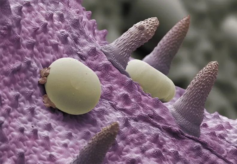
Epidermal cells

Essential oils obtained from flowers are not usually secreted by glandular hairs but merely diffuse through the cytoplasm, the cell walls, and the cuticle to the outside. The yield of essential oils from these species is generally meager. Examples include rose (Rosa spp., Rosaceae), 0.075% (w/v), acacia (Acacia spp., Fabaceae) 0.084% (w/v) and jasmine (Jasminum spp., Oleaceae) 0.04% (w/v).


Buds of numerous plant genera, such as Aesculus (Hippocastanaceae), Alnus (Betulaceae), Betula(Betulaceae), Populus (Salicaceae), Prunus (Rosaceae), and Rhamnus (Rhamnaceae), also secrete lipophilic substances, mainly flavonoid aglycones mixed with essential oils.


Secretion here occurs from epidermal cells that are covered by a cuticle. The secreted material is first eliminated into a space between the outer walls of the cells and the cuticle surrounding them, forming a blister that subsequently bursts.


Peppermint – Mentha x piperita Under Microscope | Aroma Hut Institute

Peppermint

Detail of the calyx surface of peppermint(Mentha x piperita L., Lamiaceae), showing yellow rounded sessile secretory glands and pointed non-secretory trichomes (Cryo-SEM magnified 742 times actual size). Photo © Microscopix photolibrary

Glandular trichomes

Glandular trichomes are modified epidermal hairs and can be found covering leaves, stems, and even parts of flowers such as the calyx in many plants of the Lamiaceae (Labiatae) family. These include lavender (Lavandula spp.), marjoram and oregano (Origanum spp.), and mint (Mentha spp.).


The secretory cells are attached by a single stem or basal cell in the epidermis. The outer surface of the gland is heavily cutinized. A toughened cuticle, in which no pores or perforations are present, usually wholly covers the trichome. The essential oils accumulate in subcuticular spaces and are thought to diffuse outwards through the cuticle. The glandular cells differ from normal plant cells because they have a very large nucleus and dense protoplasm that lacks a large central vacuole. There are numerous plasmodesmata (i.e., cytoplasmic threads running through cell walls, connecting the cytoplasm of adjacent cells) across the walls of the gland cells, especially between the stalk cell and the collecting cell. In the very young gland, the intracellular organization is almost identical to that of the adjacent cells, but complex changes occur as the secretory cells develop. The membrane system progressively degenerates, and in the fully developed glands, only a delicate granular cytoplasm (i.e., the living cell parts within the membrane, except the nucleus) remains.


Spike Lavender Under Microscope | Aroma Hut Institute

Spike Lavender

Detail of lower leaf surface of English lavender (Lavandula angustifolia Mill., Lamiaceae), showing a sessile secretory gland surrounded by non-secretory trichomes, which help to trap a layer of air around the leaf to reduce transpiration, or water loss (SEM, CPD magnified 931 times actual size). Photo © Microscopix photolibrary

Plant material received for these investigations was chemically fixed and critical point dried (CPD) for scanning electron microscopy (SEM) or cryogenically fixed for Cryo-SEM.

The following images and text were adapted from Secretory Structures of Aromatic and Medicinal Plants: A Review and Atlas of Micrographs. The text was written by Katerina P. Svoboda, Ph.D., and Tomas G. Svoboda. Andrew D. Syred created stunning micrograph images. Polly M. Syred designed and edited the book. Permission to use this data was made in writing to the HerbalGram.com website, where the following information was obtained.

Course Progress

Your cart is empty Continue
Shopping Cart
Subtotal:
Discount 
Discount 
View Details
- +
Sold Out